Gold »
PDB 1a52-3k3g »
3h4k »
Gold in PDB 3h4k: Crystal Structure of the Wild Type Thioredoxin Glutatione Reductase From Schistosoma Mansoni in Complex with Auranofin
Enzymatic activity of Crystal Structure of the Wild Type Thioredoxin Glutatione Reductase From Schistosoma Mansoni in Complex with Auranofin
All present enzymatic activity of Crystal Structure of the Wild Type Thioredoxin Glutatione Reductase From Schistosoma Mansoni in Complex with Auranofin:
1.6.4.5;
1.6.4.5;
Protein crystallography data
The structure of Crystal Structure of the Wild Type Thioredoxin Glutatione Reductase From Schistosoma Mansoni in Complex with Auranofin, PDB code: 3h4k
was solved by
F.Angelucci,
D.Dimastrogiovanni,
A.E.Miele,
G.Boumis,
M.Brunori,
A.Bellelli,
with X-Ray Crystallography technique. A brief refinement statistics is given in the table below:
| Resolution Low / High (Å) | 40.00 / 2.55 |
| Space group | C 1 2 1 |
| Cell size a, b, c (Å), α, β, γ (°) | 147.515, 102.166, 60.572, 90.00, 114.16, 90.00 |
| R / Rfree (%) | 23 / 25.6 |
Other elements in 3h4k:
The structure of Crystal Structure of the Wild Type Thioredoxin Glutatione Reductase From Schistosoma Mansoni in Complex with Auranofin also contains other interesting chemical elements:
| Potassium | (K) | 1 atom |
Gold Binding Sites:
The binding sites of Gold atom in the Crystal Structure of the Wild Type Thioredoxin Glutatione Reductase From Schistosoma Mansoni in Complex with Auranofin
(pdb code 3h4k). This binding sites where shown within
5.0 Angstroms radius around Gold atom.
In total 3 binding sites of Gold where determined in the Crystal Structure of the Wild Type Thioredoxin Glutatione Reductase From Schistosoma Mansoni in Complex with Auranofin, PDB code: 3h4k:
Jump to Gold binding site number: 1; 2; 3;
In total 3 binding sites of Gold where determined in the Crystal Structure of the Wild Type Thioredoxin Glutatione Reductase From Schistosoma Mansoni in Complex with Auranofin, PDB code: 3h4k:
Jump to Gold binding site number: 1; 2; 3;
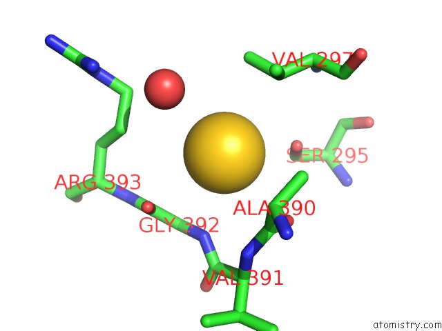
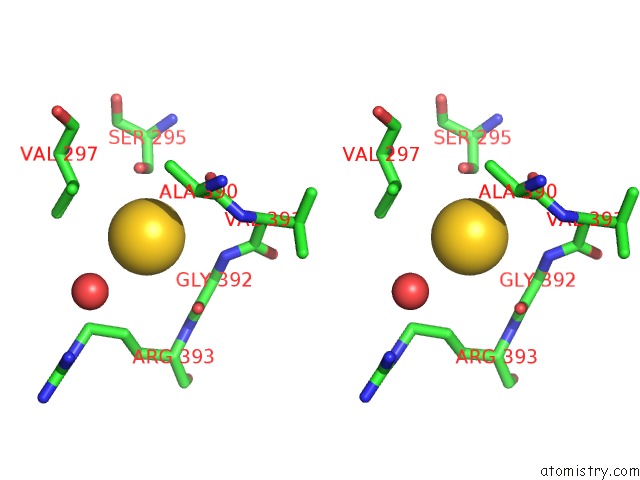
Gold binding site 1 out of 3 in 3h4k
Go back to
Gold binding site 1 out
of 3 in the Crystal Structure of the Wild Type Thioredoxin Glutatione Reductase From Schistosoma Mansoni in Complex with Auranofin
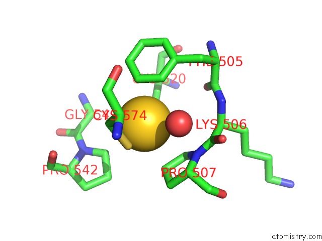
Mono view
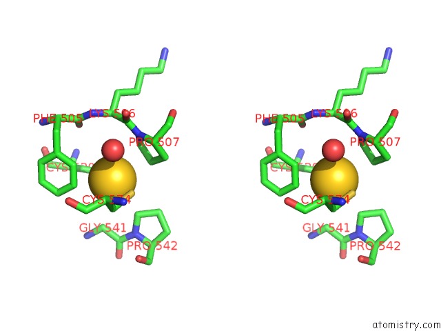
Stereo pair view
Mono view
Stereo pair view
A full contact list of Gold with other atoms in the Au binding
site number 1 of Crystal Structure of the Wild Type Thioredoxin Glutatione Reductase From Schistosoma Mansoni in Complex with Auranofin within 5.0Å range:
|
Gold binding site 2 out of 3 in 3h4k
Go back to
Gold binding site 2 out
of 3 in the Crystal Structure of the Wild Type Thioredoxin Glutatione Reductase From Schistosoma Mansoni in Complex with Auranofin
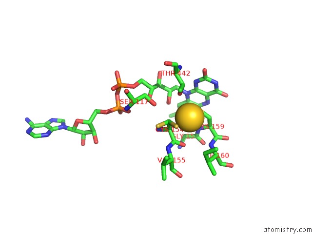
Mono view
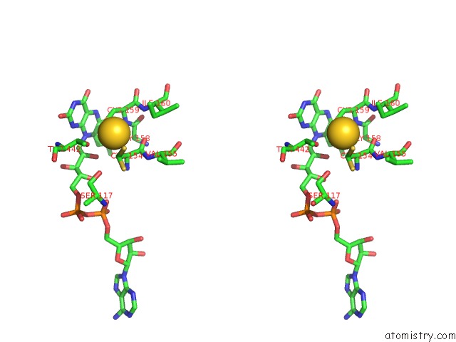
Stereo pair view
Mono view
Stereo pair view
A full contact list of Gold with other atoms in the Au binding
site number 2 of Crystal Structure of the Wild Type Thioredoxin Glutatione Reductase From Schistosoma Mansoni in Complex with Auranofin within 5.0Å range:
|
Gold binding site 3 out of 3 in 3h4k
Go back to
Gold binding site 3 out
of 3 in the Crystal Structure of the Wild Type Thioredoxin Glutatione Reductase From Schistosoma Mansoni in Complex with Auranofin
Mono view
Stereo pair view
Mono view
Stereo pair view
A full contact list of Gold with other atoms in the Au binding
site number 3 of Crystal Structure of the Wild Type Thioredoxin Glutatione Reductase From Schistosoma Mansoni in Complex with Auranofin within 5.0Å range:
|
Reference:
F.Angelucci,
A.A.Sayed,
D.L.Williams,
G.Boumis,
M.Brunori,
D.Dimastrogiovanni,
A.E.Miele,
F.Pauly,
A.Bellelli.
Inhibition of Schistosoma Mansoni Thioredoxin-Glutathione Reductase By Auranofin: Structural and Kinetic Aspects. J.Biol.Chem. V. 284 28977 2009.
ISSN: ISSN 0021-9258
PubMed: 19710012
DOI: 10.1074/JBC.M109.020701
Page generated: Mon Jul 7 01:21:55 2025
ISSN: ISSN 0021-9258
PubMed: 19710012
DOI: 10.1074/JBC.M109.020701
Last articles
Mg in 7DDQMg in 7DIY
Mg in 7DID
Mg in 7DHW
Mg in 7DIC
Mg in 7DFU
Mg in 7DFJ
Mg in 7DFK
Mg in 7DFH
Mg in 7DFG