Gold »
PDB 1a52-3k3g »
2yau »
Gold in PDB 2yau: X-Ray Structure of the Leishmania Infantum Tryopanothione Reductase in Complex with Auranofin
Enzymatic activity of X-Ray Structure of the Leishmania Infantum Tryopanothione Reductase in Complex with Auranofin
All present enzymatic activity of X-Ray Structure of the Leishmania Infantum Tryopanothione Reductase in Complex with Auranofin:
1.8.1.12;
1.8.1.12;
Protein crystallography data
The structure of X-Ray Structure of the Leishmania Infantum Tryopanothione Reductase in Complex with Auranofin, PDB code: 2yau
was solved by
P.Baiocco,
A.Ilari,
G.Colotti,
with X-Ray Crystallography technique. A brief refinement statistics is given in the table below:
| Resolution Low / High (Å) | 50.00 / 3.50 |
| Space group | P 41 |
| Cell size a, b, c (Å), α, β, γ (°) | 103.058, 103.058, 191.604, 90.00, 90.00, 90.00 |
| R / Rfree (%) | 26.704 / 31.912 |
Other elements in 2yau:
The structure of X-Ray Structure of the Leishmania Infantum Tryopanothione Reductase in Complex with Auranofin also contains other interesting chemical elements:
| Chlorine | (Cl) | 2 atoms |
Gold Binding Sites:
The binding sites of Gold atom in the X-Ray Structure of the Leishmania Infantum Tryopanothione Reductase in Complex with Auranofin
(pdb code 2yau). This binding sites where shown within
5.0 Angstroms radius around Gold atom.
In total 2 binding sites of Gold where determined in the X-Ray Structure of the Leishmania Infantum Tryopanothione Reductase in Complex with Auranofin, PDB code: 2yau:
Jump to Gold binding site number: 1; 2;
In total 2 binding sites of Gold where determined in the X-Ray Structure of the Leishmania Infantum Tryopanothione Reductase in Complex with Auranofin, PDB code: 2yau:
Jump to Gold binding site number: 1; 2;
Gold binding site 1 out of 2 in 2yau
Go back to
Gold binding site 1 out
of 2 in the X-Ray Structure of the Leishmania Infantum Tryopanothione Reductase in Complex with Auranofin
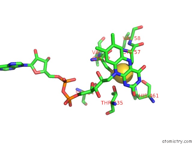
Mono view
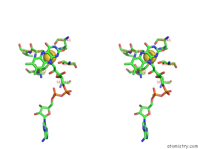
Stereo pair view
Mono view
Stereo pair view
A full contact list of Gold with other atoms in the Au binding
site number 1 of X-Ray Structure of the Leishmania Infantum Tryopanothione Reductase in Complex with Auranofin within 5.0Å range:
|
Gold binding site 2 out of 2 in 2yau
Go back to
Gold binding site 2 out
of 2 in the X-Ray Structure of the Leishmania Infantum Tryopanothione Reductase in Complex with Auranofin
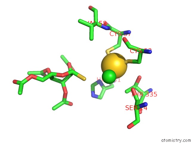
Mono view
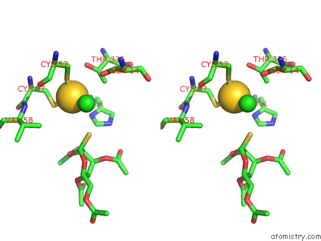
Stereo pair view
Mono view
Stereo pair view
A full contact list of Gold with other atoms in the Au binding
site number 2 of X-Ray Structure of the Leishmania Infantum Tryopanothione Reductase in Complex with Auranofin within 5.0Å range:
|
Reference:
A.Ilari,
P.Baiocco,
L.Messori,
A.Fiorillo,
A.Boffi,
M.Gramiccia,
T.Di Muccio,
G.Colotti.
A Gold-Containing Drug Against Parasitic Polyamine Metabolism: the X-Ray Structure of Trypanothione Reductase From Leishmania Infantum in Complex with Auranofin Reveals A Dual Mechanism of Enzyme Inhibition. Amino Acids V. 42 803 2012.
ISSN: ISSN 0939-4451
PubMed: 21833767
DOI: 10.1007/S00726-011-0997-9
Page generated: Mon Jul 7 01:21:26 2025
ISSN: ISSN 0939-4451
PubMed: 21833767
DOI: 10.1007/S00726-011-0997-9
Last articles
K in 5ZYAK in 5ZOF
K in 5ZOE
K in 5ZE2
K in 5ZE1
K in 5YSH
K in 5YRV
K in 5YRT
K in 5ZE0
K in 5ZDZ