Gold »
PDB 5jjo-7pvj »
6seu »
Gold in PDB 6seu: X-Ray Structure of the Gold/Lysozyme Adduct Formed Upon 21H Exposure of Protein Crystals to Compound 2
Enzymatic activity of X-Ray Structure of the Gold/Lysozyme Adduct Formed Upon 21H Exposure of Protein Crystals to Compound 2
All present enzymatic activity of X-Ray Structure of the Gold/Lysozyme Adduct Formed Upon 21H Exposure of Protein Crystals to Compound 2:
3.2.1.17;
3.2.1.17;
Protein crystallography data
The structure of X-Ray Structure of the Gold/Lysozyme Adduct Formed Upon 21H Exposure of Protein Crystals to Compound 2, PDB code: 6seu
was solved by
G.Ferraro,
A.Giorgio,
A.Merlino,
with X-Ray Crystallography technique. A brief refinement statistics is given in the table below:
| Resolution Low / High (Å) | 34.73 / 1.95 |
| Space group | P 43 21 2 |
| Cell size a, b, c (Å), α, β, γ (°) | 76.953, 76.953, 38.889, 90.00, 90.00, 90.00 |
| R / Rfree (%) | 19.2 / 26 |
Gold Binding Sites:
The binding sites of Gold atom in the X-Ray Structure of the Gold/Lysozyme Adduct Formed Upon 21H Exposure of Protein Crystals to Compound 2
(pdb code 6seu). This binding sites where shown within
5.0 Angstroms radius around Gold atom.
In total 4 binding sites of Gold where determined in the X-Ray Structure of the Gold/Lysozyme Adduct Formed Upon 21H Exposure of Protein Crystals to Compound 2, PDB code: 6seu:
Jump to Gold binding site number: 1; 2; 3; 4;
In total 4 binding sites of Gold where determined in the X-Ray Structure of the Gold/Lysozyme Adduct Formed Upon 21H Exposure of Protein Crystals to Compound 2, PDB code: 6seu:
Jump to Gold binding site number: 1; 2; 3; 4;
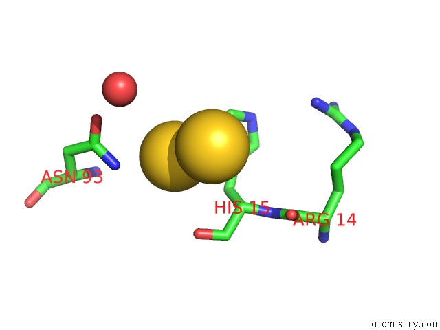
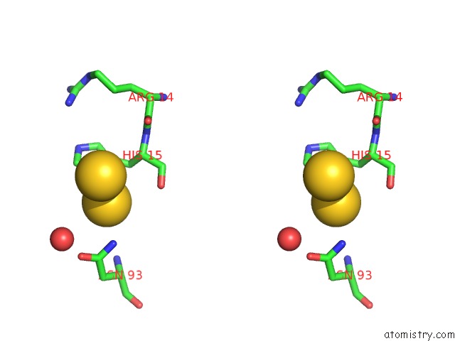
Gold binding site 1 out of 4 in 6seu
Go back to
Gold binding site 1 out
of 4 in the X-Ray Structure of the Gold/Lysozyme Adduct Formed Upon 21H Exposure of Protein Crystals to Compound 2
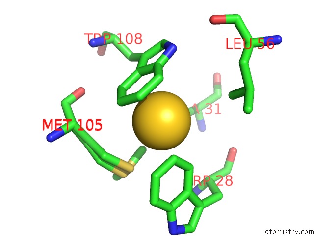
Mono view
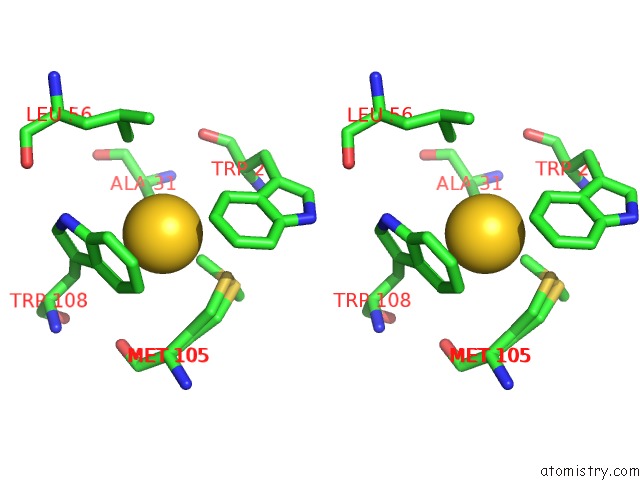
Stereo pair view
Mono view
Stereo pair view
A full contact list of Gold with other atoms in the Au binding
site number 1 of X-Ray Structure of the Gold/Lysozyme Adduct Formed Upon 21H Exposure of Protein Crystals to Compound 2 within 5.0Å range:
|
Gold binding site 2 out of 4 in 6seu
Go back to
Gold binding site 2 out
of 4 in the X-Ray Structure of the Gold/Lysozyme Adduct Formed Upon 21H Exposure of Protein Crystals to Compound 2
Mono view
Stereo pair view
Mono view
Stereo pair view
A full contact list of Gold with other atoms in the Au binding
site number 2 of X-Ray Structure of the Gold/Lysozyme Adduct Formed Upon 21H Exposure of Protein Crystals to Compound 2 within 5.0Å range:
|
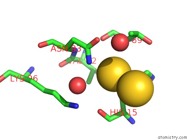
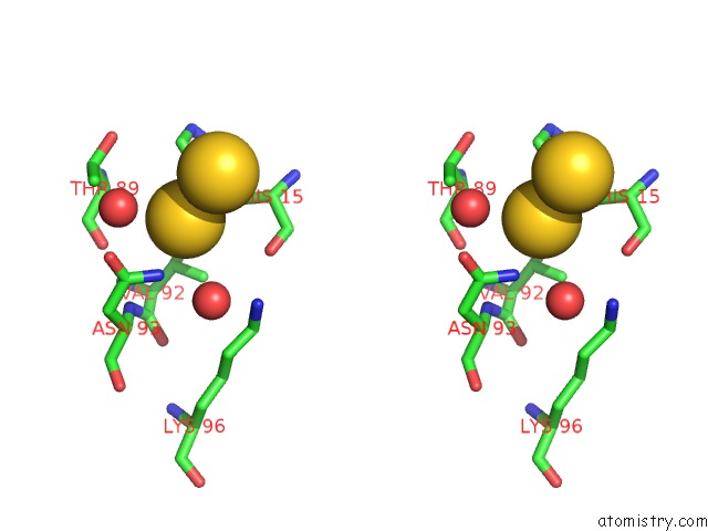
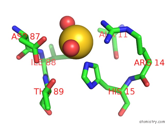
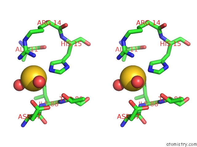
Gold binding site 3 out of 4 in 6seu
Go back to
Gold binding site 3 out
of 4 in the X-Ray Structure of the Gold/Lysozyme Adduct Formed Upon 21H Exposure of Protein Crystals to Compound 2
Mono view
Stereo pair view
Mono view
Stereo pair view
A full contact list of Gold with other atoms in the Au binding
site number 3 of X-Ray Structure of the Gold/Lysozyme Adduct Formed Upon 21H Exposure of Protein Crystals to Compound 2 within 5.0Å range:
|
Gold binding site 4 out of 4 in 6seu
Go back to
Gold binding site 4 out
of 4 in the X-Ray Structure of the Gold/Lysozyme Adduct Formed Upon 21H Exposure of Protein Crystals to Compound 2
Mono view
Stereo pair view
Mono view
Stereo pair view
A full contact list of Gold with other atoms in the Au binding
site number 4 of X-Ray Structure of the Gold/Lysozyme Adduct Formed Upon 21H Exposure of Protein Crystals to Compound 2 within 5.0Å range:
|
Reference:
G.Ferraro,
A.Giorgio,
A.M.Mansour,
A.Merlino.
Protein-Mediated Disproportionation of Au(I): Insights From the Structures of Adducts of Au(III) Compounds Bearing N,N-Pyridylbenzimidazole Derivatives with Lysozyme. Dalton Trans V. 48 14027 2019.
ISSN: ESSN 1477-9234
PubMed: 31490509
DOI: 10.1039/C9DT02729G
Page generated: Mon Jul 7 01:40:07 2025
ISSN: ESSN 1477-9234
PubMed: 31490509
DOI: 10.1039/C9DT02729G
Last articles
Cl in 5CLMCl in 5CKX
Cl in 5CLS
Cl in 5CKV
Cl in 5CKL
Cl in 5CLK
Cl in 5CJ3
Cl in 5CJJ
Cl in 5CJ6
Cl in 5CHB