Gold »
PDB 5jjo-7pvj »
6i9y »
Gold in PDB 6i9y: The 2.14 A X-Ray Crystal Structure of Sporosarcina Pasteurii Urease in Complex with Au(I) Ions
Enzymatic activity of The 2.14 A X-Ray Crystal Structure of Sporosarcina Pasteurii Urease in Complex with Au(I) Ions
All present enzymatic activity of The 2.14 A X-Ray Crystal Structure of Sporosarcina Pasteurii Urease in Complex with Au(I) Ions:
3.5.1.5;
3.5.1.5;
Protein crystallography data
The structure of The 2.14 A X-Ray Crystal Structure of Sporosarcina Pasteurii Urease in Complex with Au(I) Ions, PDB code: 6i9y
was solved by
L.Mazzei,
M.Cianci,
S.Ciurli,
with X-Ray Crystallography technique. A brief refinement statistics is given in the table below:
| Resolution Low / High (Å) | 45.80 / 2.14 |
| Space group | P 63 2 2 |
| Cell size a, b, c (Å), α, β, γ (°) | 132.291, 132.291, 190.435, 90.00, 90.00, 120.00 |
| R / Rfree (%) | 17.1 / 20.8 |
Other elements in 6i9y:
The structure of The 2.14 A X-Ray Crystal Structure of Sporosarcina Pasteurii Urease in Complex with Au(I) Ions also contains other interesting chemical elements:
| Nickel | (Ni) | 2 atoms |
Gold Binding Sites:
The binding sites of Gold atom in the The 2.14 A X-Ray Crystal Structure of Sporosarcina Pasteurii Urease in Complex with Au(I) Ions
(pdb code 6i9y). This binding sites where shown within
5.0 Angstroms radius around Gold atom.
In total 3 binding sites of Gold where determined in the The 2.14 A X-Ray Crystal Structure of Sporosarcina Pasteurii Urease in Complex with Au(I) Ions, PDB code: 6i9y:
Jump to Gold binding site number: 1; 2; 3;
In total 3 binding sites of Gold where determined in the The 2.14 A X-Ray Crystal Structure of Sporosarcina Pasteurii Urease in Complex with Au(I) Ions, PDB code: 6i9y:
Jump to Gold binding site number: 1; 2; 3;
Gold binding site 1 out of 3 in 6i9y
Go back to
Gold binding site 1 out
of 3 in the The 2.14 A X-Ray Crystal Structure of Sporosarcina Pasteurii Urease in Complex with Au(I) Ions
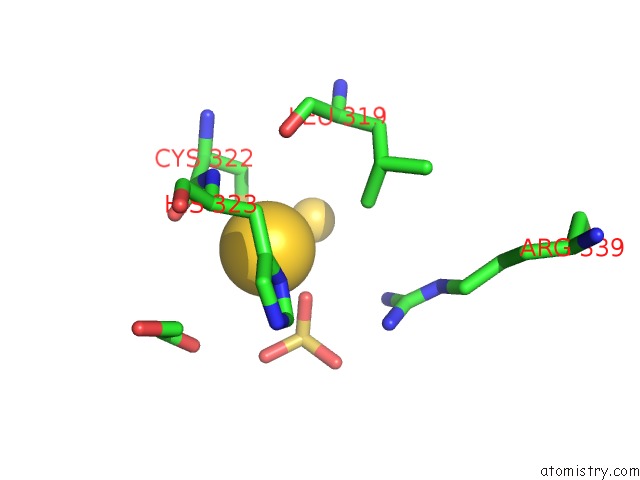
Mono view
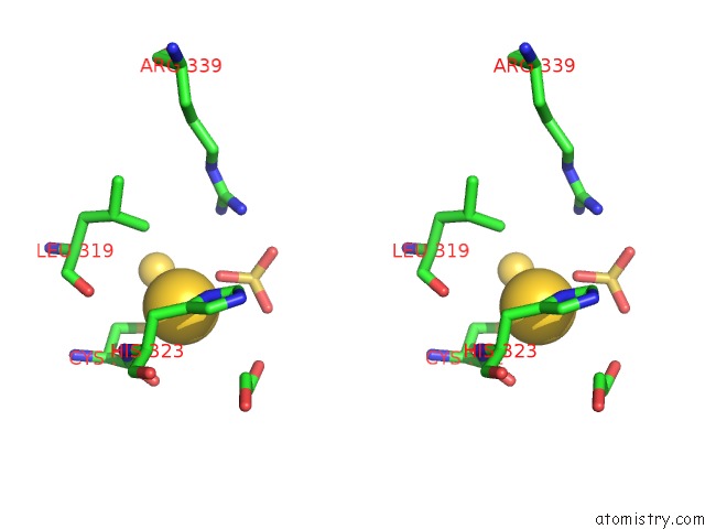
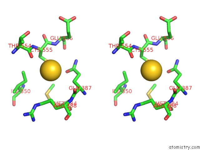
Stereo pair view
Mono view
Stereo pair view
A full contact list of Gold with other atoms in the Au binding
site number 1 of The 2.14 A X-Ray Crystal Structure of Sporosarcina Pasteurii Urease in Complex with Au(I) Ions within 5.0Å range:
|
Gold binding site 2 out of 3 in 6i9y
Go back to
Gold binding site 2 out
of 3 in the The 2.14 A X-Ray Crystal Structure of Sporosarcina Pasteurii Urease in Complex with Au(I) Ions
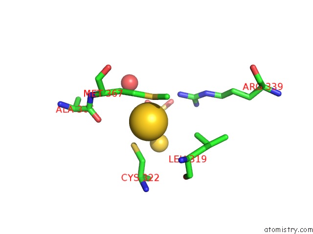
Mono view
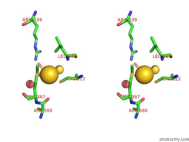
Stereo pair view
Mono view
Stereo pair view
A full contact list of Gold with other atoms in the Au binding
site number 2 of The 2.14 A X-Ray Crystal Structure of Sporosarcina Pasteurii Urease in Complex with Au(I) Ions within 5.0Å range:
|
Gold binding site 3 out of 3 in 6i9y
Go back to
Gold binding site 3 out
of 3 in the The 2.14 A X-Ray Crystal Structure of Sporosarcina Pasteurii Urease in Complex with Au(I) Ions

Mono view
Stereo pair view
Mono view
Stereo pair view
A full contact list of Gold with other atoms in the Au binding
site number 3 of The 2.14 A X-Ray Crystal Structure of Sporosarcina Pasteurii Urease in Complex with Au(I) Ions within 5.0Å range:
|
Reference:
L.Mazzei,
M.N.Wenzel,
M.Cianci,
M.Palombo,
A.Casini,
S.Ciurli.
Inhibition Mechanism of Urease By Au(III) Compounds Unveiled By X-Ray Diffraction Analysis. Acs Med.Chem.Lett. V. 10 564 2019.
ISSN: ISSN 1948-5875
PubMed: 30996797
DOI: 10.1021/ACSMEDCHEMLETT.8B00585
Page generated: Mon Jul 7 01:38:49 2025
ISSN: ISSN 1948-5875
PubMed: 30996797
DOI: 10.1021/ACSMEDCHEMLETT.8B00585
Last articles
Fe in 2YXOFe in 2YRS
Fe in 2YXC
Fe in 2YNM
Fe in 2YVJ
Fe in 2YP1
Fe in 2YU2
Fe in 2YU1
Fe in 2YQB
Fe in 2YOO