Gold »
PDB 1a52-3k3g »
2yau »
Gold in PDB 2yau: X-Ray Structure of the Leishmania Infantum Tryopanothione Reductase in Complex with Auranofin
Enzymatic activity of X-Ray Structure of the Leishmania Infantum Tryopanothione Reductase in Complex with Auranofin
All present enzymatic activity of X-Ray Structure of the Leishmania Infantum Tryopanothione Reductase in Complex with Auranofin:
1.8.1.12;
1.8.1.12;
Protein crystallography data
The structure of X-Ray Structure of the Leishmania Infantum Tryopanothione Reductase in Complex with Auranofin, PDB code: 2yau
was solved by
P.Baiocco,
A.Ilari,
G.Colotti,
with X-Ray Crystallography technique. A brief refinement statistics is given in the table below:
| Resolution Low / High (Å) | 50.00 / 3.50 |
| Space group | P 41 |
| Cell size a, b, c (Å), α, β, γ (°) | 103.058, 103.058, 191.604, 90.00, 90.00, 90.00 |
| R / Rfree (%) | 26.704 / 31.912 |
Other elements in 2yau:
The structure of X-Ray Structure of the Leishmania Infantum Tryopanothione Reductase in Complex with Auranofin also contains other interesting chemical elements:
| Chlorine | (Cl) | 2 atoms |
Gold Binding Sites:
The binding sites of Gold atom in the X-Ray Structure of the Leishmania Infantum Tryopanothione Reductase in Complex with Auranofin
(pdb code 2yau). This binding sites where shown within
5.0 Angstroms radius around Gold atom.
In total 2 binding sites of Gold where determined in the X-Ray Structure of the Leishmania Infantum Tryopanothione Reductase in Complex with Auranofin, PDB code: 2yau:
Jump to Gold binding site number: 1; 2;
In total 2 binding sites of Gold where determined in the X-Ray Structure of the Leishmania Infantum Tryopanothione Reductase in Complex with Auranofin, PDB code: 2yau:
Jump to Gold binding site number: 1; 2;
Gold binding site 1 out of 2 in 2yau
Go back to
Gold binding site 1 out
of 2 in the X-Ray Structure of the Leishmania Infantum Tryopanothione Reductase in Complex with Auranofin
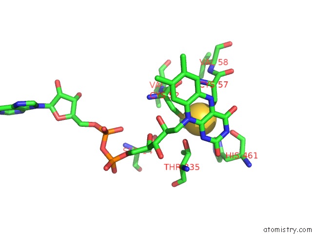
Mono view
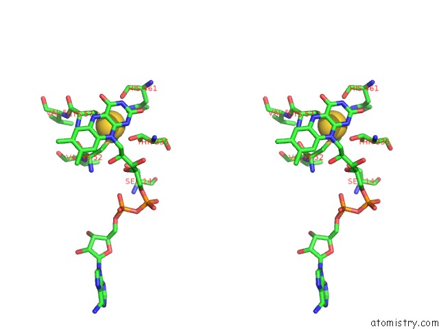
Stereo pair view
Mono view
Stereo pair view
A full contact list of Gold with other atoms in the Au binding
site number 1 of X-Ray Structure of the Leishmania Infantum Tryopanothione Reductase in Complex with Auranofin within 5.0Å range:
|
Gold binding site 2 out of 2 in 2yau
Go back to
Gold binding site 2 out
of 2 in the X-Ray Structure of the Leishmania Infantum Tryopanothione Reductase in Complex with Auranofin
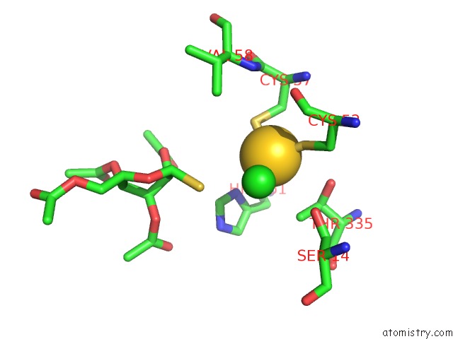
Mono view
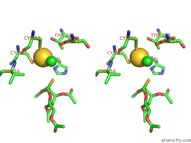
Stereo pair view
Mono view
Stereo pair view
A full contact list of Gold with other atoms in the Au binding
site number 2 of X-Ray Structure of the Leishmania Infantum Tryopanothione Reductase in Complex with Auranofin within 5.0Å range:
|
Reference:
A.Ilari,
P.Baiocco,
L.Messori,
A.Fiorillo,
A.Boffi,
M.Gramiccia,
T.Di Muccio,
G.Colotti.
A Gold-Containing Drug Against Parasitic Polyamine Metabolism: the X-Ray Structure of Trypanothione Reductase From Leishmania Infantum in Complex with Auranofin Reveals A Dual Mechanism of Enzyme Inhibition. Amino Acids V. 42 803 2012.
ISSN: ISSN 0939-4451
PubMed: 21833767
DOI: 10.1007/S00726-011-0997-9
Page generated: Wed Jul 10 14:13:47 2024
ISSN: ISSN 0939-4451
PubMed: 21833767
DOI: 10.1007/S00726-011-0997-9
Last articles
Zn in 9MJ5Zn in 9HNW
Zn in 9G0L
Zn in 9FNE
Zn in 9DZN
Zn in 9E0I
Zn in 9D32
Zn in 9DAK
Zn in 8ZXC
Zn in 8ZUF