Gold »
PDB 1a52-3k3g »
1n8n »
Gold in PDB 1n8n: Crystal Structure of the AU3+ Complex of Apha Class B Acid Phosphatase/Phosphotransferase From E. Coli at 1.69 A Resolution
Enzymatic activity of Crystal Structure of the AU3+ Complex of Apha Class B Acid Phosphatase/Phosphotransferase From E. Coli at 1.69 A Resolution
All present enzymatic activity of Crystal Structure of the AU3+ Complex of Apha Class B Acid Phosphatase/Phosphotransferase From E. Coli at 1.69 A Resolution:
3.1.3.2;
3.1.3.2;
Protein crystallography data
The structure of Crystal Structure of the AU3+ Complex of Apha Class B Acid Phosphatase/Phosphotransferase From E. Coli at 1.69 A Resolution, PDB code: 1n8n
was solved by
V.Calderone,
C.Forleo,
M.Benvenuti,
G.M.Rossolini,
M.C.Thaller,
S.Mangani,
with X-Ray Crystallography technique. A brief refinement statistics is given in the table below:
| Resolution Low / High (Å) | 46.13 / 1.69 |
| Space group | I 2 2 2 |
| Cell size a, b, c (Å), α, β, γ (°) | 49.279, 92.457, 138.182, 90.00, 90.00, 90.00 |
| R / Rfree (%) | 17.8 / 20.7 |
Gold Binding Sites:
The binding sites of Gold atom in the Crystal Structure of the AU3+ Complex of Apha Class B Acid Phosphatase/Phosphotransferase From E. Coli at 1.69 A Resolution
(pdb code 1n8n). This binding sites where shown within
5.0 Angstroms radius around Gold atom.
In total only one binding site of Gold was determined in the Crystal Structure of the AU3+ Complex of Apha Class B Acid Phosphatase/Phosphotransferase From E. Coli at 1.69 A Resolution, PDB code: 1n8n:
In total only one binding site of Gold was determined in the Crystal Structure of the AU3+ Complex of Apha Class B Acid Phosphatase/Phosphotransferase From E. Coli at 1.69 A Resolution, PDB code: 1n8n:
Gold binding site 1 out of 1 in 1n8n
Go back to
Gold binding site 1 out
of 1 in the Crystal Structure of the AU3+ Complex of Apha Class B Acid Phosphatase/Phosphotransferase From E. Coli at 1.69 A Resolution
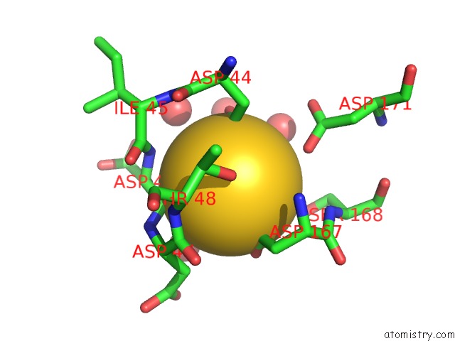
Mono view
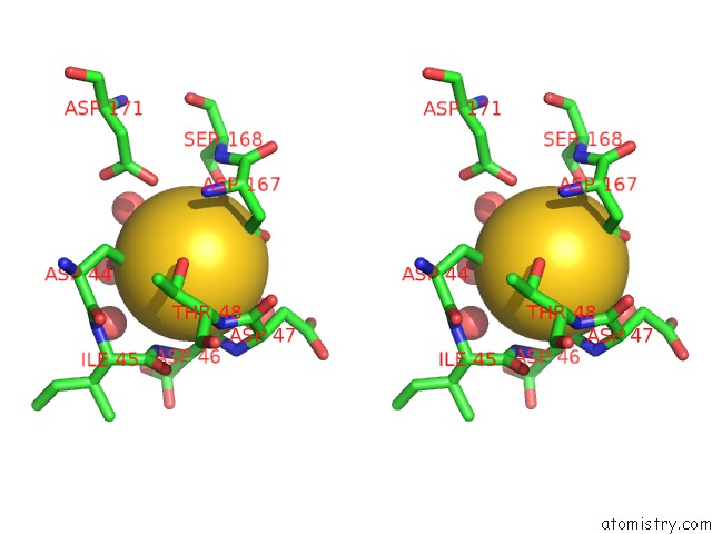
Stereo pair view
Mono view
Stereo pair view
A full contact list of Gold with other atoms in the Au binding
site number 1 of Crystal Structure of the AU3+ Complex of Apha Class B Acid Phosphatase/Phosphotransferase From E. Coli at 1.69 A Resolution within 5.0Å range:
|
Reference:
V.Calderone,
C.Forleo,
M.Benvenuti,
M.C.Thaller,
G.M.Rossolini,
S.Mangani.
The First Structure of A Bacterial Class B Acid Phosphatase Reveals Further Structural Heterogeneity Among Phosphatases of the Haloacid Dehalogenase Fold. J.Mol.Biol. V. 335 761 2004.
ISSN: ISSN 0022-2836
PubMed: 14687572
DOI: 10.1016/J.JMB.2003.10.050
Page generated: Mon Jul 7 01:18:37 2025
ISSN: ISSN 0022-2836
PubMed: 14687572
DOI: 10.1016/J.JMB.2003.10.050
Last articles
F in 4OHAF in 4OH5
F in 4OH6
F in 4OGH
F in 4OFB
F in 4OG6
F in 4ODE
F in 4O9S
F in 4OD0
F in 4OCZ