Gold »
PDB 1a52-3k3g »
1a79 »
Gold in PDB 1a79: Crystal Structure of the Trna Splicing Endonuclease From Methanococcus Jannaschii
Protein crystallography data
The structure of Crystal Structure of the Trna Splicing Endonuclease From Methanococcus Jannaschii, PDB code: 1a79
was solved by
H.Li,
C.R.Trotta,
J.N.Abelson,
with X-Ray Crystallography technique. A brief refinement statistics is given in the table below:
| Resolution Low / High (Å) | 50.00 / 2.28 |
| Space group | P 21 21 21 |
| Cell size a, b, c (Å), α, β, γ (°) | 61.950, 80.040, 193.590, 90.00, 90.00, 90.00 |
| R / Rfree (%) | 20 / 26.7 |
Gold Binding Sites:
The binding sites of Gold atom in the Crystal Structure of the Trna Splicing Endonuclease From Methanococcus Jannaschii
(pdb code 1a79). This binding sites where shown within
5.0 Angstroms radius around Gold atom.
In total 4 binding sites of Gold where determined in the Crystal Structure of the Trna Splicing Endonuclease From Methanococcus Jannaschii, PDB code: 1a79:
Jump to Gold binding site number: 1; 2; 3; 4;
In total 4 binding sites of Gold where determined in the Crystal Structure of the Trna Splicing Endonuclease From Methanococcus Jannaschii, PDB code: 1a79:
Jump to Gold binding site number: 1; 2; 3; 4;
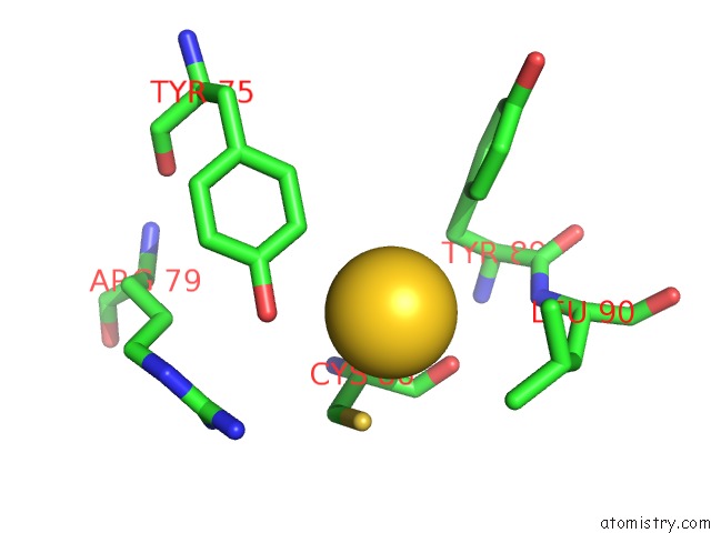
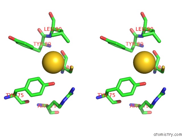
Gold binding site 1 out of 4 in 1a79
Go back to
Gold binding site 1 out
of 4 in the Crystal Structure of the Trna Splicing Endonuclease From Methanococcus Jannaschii
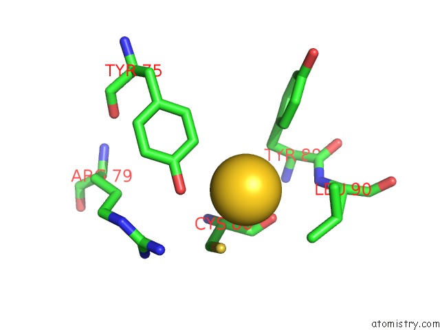
Mono view
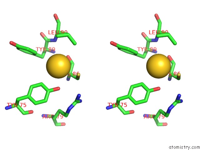
Stereo pair view
Mono view
Stereo pair view
A full contact list of Gold with other atoms in the Au binding
site number 1 of Crystal Structure of the Trna Splicing Endonuclease From Methanococcus Jannaschii within 5.0Å range:
|
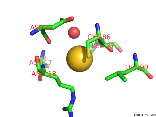
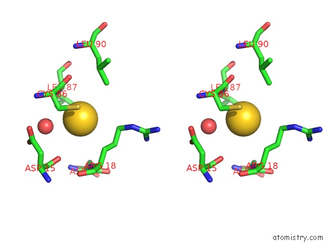
Gold binding site 2 out of 4 in 1a79
Go back to
Gold binding site 2 out
of 4 in the Crystal Structure of the Trna Splicing Endonuclease From Methanococcus Jannaschii
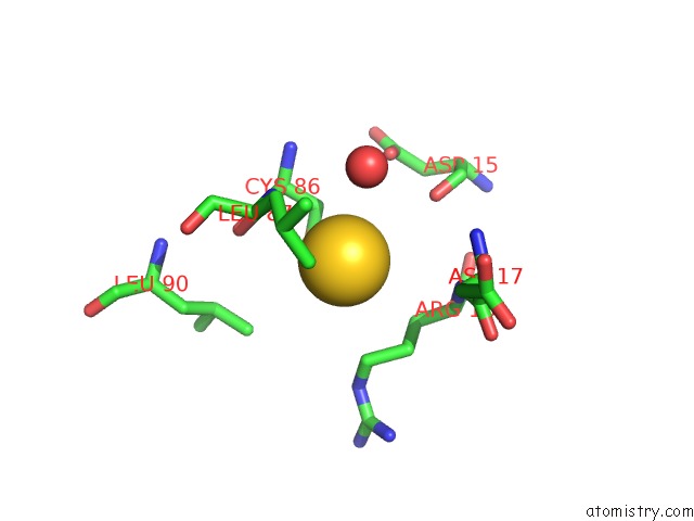
Mono view
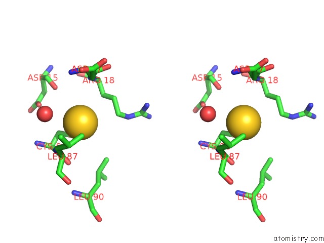
Stereo pair view
Mono view
Stereo pair view
A full contact list of Gold with other atoms in the Au binding
site number 2 of Crystal Structure of the Trna Splicing Endonuclease From Methanococcus Jannaschii within 5.0Å range:
|
Gold binding site 3 out of 4 in 1a79
Go back to
Gold binding site 3 out
of 4 in the Crystal Structure of the Trna Splicing Endonuclease From Methanococcus Jannaschii
Mono view
Stereo pair view
Mono view
Stereo pair view
A full contact list of Gold with other atoms in the Au binding
site number 3 of Crystal Structure of the Trna Splicing Endonuclease From Methanococcus Jannaschii within 5.0Å range:
|
Gold binding site 4 out of 4 in 1a79
Go back to
Gold binding site 4 out
of 4 in the Crystal Structure of the Trna Splicing Endonuclease From Methanococcus Jannaschii
Mono view
Stereo pair view
Mono view
Stereo pair view
A full contact list of Gold with other atoms in the Au binding
site number 4 of Crystal Structure of the Trna Splicing Endonuclease From Methanococcus Jannaschii within 5.0Å range:
|
Reference:
H.Li,
C.R.Trotta,
J.Abelson.
Crystal Structure and Evolution of A Transfer Rna Splicing Enzyme. Science V. 280 279 1998.
ISSN: ISSN 0036-8075
PubMed: 9535656
DOI: 10.1126/SCIENCE.280.5361.279
Page generated: Wed Jul 10 14:08:51 2024
ISSN: ISSN 0036-8075
PubMed: 9535656
DOI: 10.1126/SCIENCE.280.5361.279
Last articles
Zn in 9J0NZn in 9J0O
Zn in 9J0P
Zn in 9FJX
Zn in 9EKB
Zn in 9C0F
Zn in 9CAH
Zn in 9CH0
Zn in 9CH3
Zn in 9CH1